Allergy is an excessive and inappropriate response of the immune system to a normally harmless substance (allergen), resulting in the release of inflammatory mediators and characteristic symptoms.
In the first part, we will look at a controversy surrounding the lack of a cutaneous barrier in atopic skin in dogs: is this lesion secondary to inflammation, or does it pre-exist? Dr Anne Roussel, a specialist in dermatology, will clarify this question by summarising a study by Thierry Olivry et al. published in Veterinary Dermatology.
In the second part of our Newsletter, we present a review of the scientific literature on the most common allergies in dogs and cats, with a particular focus on treatment with allergen-specific immunotherapy.
Enjoy your reading!
 Quote of the month: "You can't run and scratch your feet at the same time".
Malian proverb
The expert summary: transient and reversible reduction of filaggrin degradation products in the stratum corneum after allergic challenge in atopic dogs experimentally sensitised to mites.
Transient and reversible reduction of stratum corneum filaggrin degradation products after allergen challenge in experimentally mite-sensitised atopic dogs. Thierry Olivry, Judy S. Paps and Nicolas Amalric. Vet Dermatol 2022; 33: 62-e20
Summary by: Dr Anne Roussel, DMV, specialist in veterinary dermatology (Dipl. ECVD), CES in dermatology. 06000 Nice
Introduction :There is growing evidence that the skin barrier plays a central role in the pathogenesis of atopic dermatitis in humans and dogs. Researchers are currently working to determine whether these skin barrier abnormalities are inherent in a primary genetic defect in the epithelial lining or are induced directly by Th2-type allergic inflammation.
Aims of the study :The aim of the study was to observe whether an allergic challenge reduces the production of natural moisturising factor (NMF) containing the main degradation products of filaggrin (FLG).
Focus on filaggrin : FLG contributes to the architecture of the stratum corneum by helping to flatten corneocytes and facilitating the migration of lamellar bodies (Figure 2).
FLG is broken down at the skin's surface into amino acids, which contribute to the formation of natural moisturising factor (NMF).
When it is broken down, FLG releases histidine, which helps to acidify the skin after being converted into urocanic acid. The acidic pH of the skin inhibits bacterial development and is essential for skin differentiation and cornification.
In humans, over 40 different mutations are currently recognised in patients suffering from ichthyosis vulgaris and/or AD (18% to 48% of atopic patients are thought to have a mutation in the gene encoding FLG).
The mutation is associated with an increased risk of developing eczema, food allergy, asthma and allergic rhinitis. However, not all individuals with an FLG mutation have an allergic phenotype.
In dogs, immunohistochemical (IHC) studies show that abnormalities in FLG expression are frequent in atopic dogs, but the clinical involvement of FLG in the pathogenesis of atopic dermatitis is still poorly understood (from a quantitative point of view, there does not appear to be an inverse correlation between the severity of CAD and FLG expression) and no mutation has yet been clearly identified in this species.
Materials and methods : Epicutaneous allergic stimulation with Dermatophagoides farinae (Df) was performed on four experimentally sensitised Beagle dogs. The patch test was applied to two sites. Several skin samples were taken at both sites on days (D)1, D2, D3, D7 and D28, after the allergic challenge. The components of the natural hydration factor were assessed by liquid chromatography and tandem mass spectrometry (LC/MS-MS).
Results :The allergic challenge test induced moderate skin lesions at the application site. Minor erythema was noted at the control application site.
Allergic stimulation leads to a significant reduction in total NMF and its components.
These observations apply both to the site where the allergic challenge was carried out and, with a slight time lag, to the site where only the control vehicle (mineral oil) was applied.
Lesion scores decreased on the seventh day and NMF concentrations increased again on the twenty-eighth day. The more pronounced the skin lesions, the lower the total concentrations of NMF.
Conclusions and discussion: In this experimental model, a single epicutaneous allergic challenge produced skin lesions, as well as a transient and reversible decrease in total NMF and its components. Similar observations were made with skin ceramides and skin microbiota. These changes were reversible once the inflammation had been brought under control, suggesting anomalies in the cutaneous barrier secondary to allergic inflammation. The author suggests a reduction in the activity of enzymes involved in the degradation of filaggrin to NMF rather than an absence of expression.
In addition, the changes observed at the challenge site and at a distance in the control area point to a systemic allergic inflammatory reaction and not simply to a condition confined to the skin.
It is important to bear in mind the very small sample of dogs in this study and the fact that this is a model of dogs experimentally sensitised to dust mites.
Finally, it is important to remember the diversity of dog breeds and the possible individual variability of barrier defects associated with atopic dermatitis, particularly regarding filaggrin.
Literature review: what are the most common allergies in dogs and cats?
Allergy to flea bites (DAPP) is one of the most common allergies (allergy to the bites of other insects, including mosquitoes, is much rarer1,2 ).
Food allergies, which are also common, are, in the vast majority of cases, due to animal proteins such as beef, dairy products, chicken and lamb (ahead of eggs, fish and pork) and sometimes to plant proteins (wheat, soya, rice and maize)3 .
Contact allergies, resulting from an antigenic reaction to an allergenic agent, are difficult to differentiate from contact dermatitis due to a non-specific irritant agent4 (in both cases, it may be, for example, topical medicines, shampoos, cleaning agents, insecticides, plants, metals or textiles).
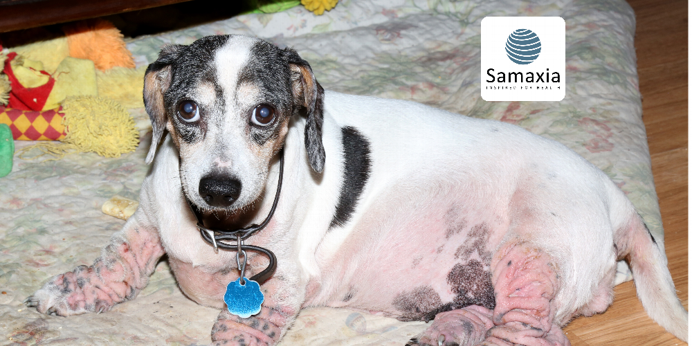
Finally, atopic dermatitis is a well-known genetic allergy in dogs, involving IgE antibodies and caused by environmental allergens (e.g. house dust mites, moulds or pollens). The skin barrier is generally damaged, facilitating contact between the cutaneous immune system and environmental allergens5,6 .
Does atopic dermatitis exist in cats?
This is a question that divides the scientific community! The cat is not a small dog, as is once again proven. In cats, the term "feline atopic syndrome" is often used to describe allergies that are not caused by flea saliva or food. However, recent studies seem to indicate the presence of IgE in the pathogenesis of the dermatitis-asthma-enteritis triad, which would justify considering cats to be suffering from atopic diseases6,7 .
Focus on desensitisation: The aim of ASIT (Allergen-Specific Immunotherapy) is to reduce the patient's immune response to the agents responsible for his allergy. Once the allergens have been identified based on allergy tests and the prevalence of allergens in the region where the animal lives and they are administered in increasing concentrations over an induction period. Injections are spaced out when the maintenance dose is reached. The allergen dose and frequency of administration are adapted to each animal individually based on the clinical results obtained8,9,10.
Effectiveness of treatment: In 70 to 80% of allergic dogs and cats, ASIT reduces symptoms by at least 50% over 9 to 18 months11. Desensitisation does not therefore guarantee a cure (although one study found complete remission in 5% of dogs suffering from atopic dermatitis12), but by reducing the intensity of symptoms, it significantly improves the animal's quality of life13.
Administration: The traditional approach is subcutaneous injection (SCIT). Alternative routes of administration are being explored but require further study. For example, the sublingual route (SLIT) has produced good results in animals that did not respond or had side effects following injections14, and the intra lymphatic route (ILIT) is thought to produce a more rapid and targeted immune response 15,16.
How long does the treatment last? ASIT should ideally be continued for at least 9 to 12 months before results are assessed. Some patients will require lifelong therapy, while others will enjoy prolonged remission for years after stopping ASIT11 .
Should I be concerned about side effects? Exacerbated pruritus or irritation at the injection site may occur in a small number of cases, requiring adjustment (not discontinuation) of treatment. Systemic reactions have been reported in approximately 1% of dogs and include weakness, depression, drowsiness, panting, diarrhoea, vomiting, urticaria, angioedema and anaphylaxis, however, in general, ASIT is considered a safe and effective method of desensitisation for allergic animals10 .
References
Allergies to insect bites
Mueller RS, Janda J, Jensen-Jarolim E, Rhyner C, Marti E. Allergens in veterina ry medicine. Allergy. 2016 Jan;71(1):27-35. doi: 10.1111/all.12726. Epub 2015 Sep 11. PMID: 26280544; PMCID: PMC4716287.
Diesel A. Cutaneous Hypersensitivity Dermatoses in the Feline Patient: A Review of Allergic Skin Disease in Cats. Vet Sci. 2017 May 9;4(2):25. doi: 10.3390/vetsci4020025. PMID: 29056684; PMCID: PMC5606602.
Food allergies and contact allergies
Tiffany S, Parr JM, Templeman J, Shoveller AK, Manjos R, Yu A, Verbrugghe A. Assessment of dog owners' knowledge relating to the diagnosis and treatment of canine food allergies. Can Vet J. 2019 Mar;60(3):268-274. PMID: 30872849; PMCID: PMC6380261.
Walder EJ, Conroy JD. Contact Dermatitis in Dogs and Cats: Pathogenesis, Histopathology, Experimental Induction and Case Reports. Vet Dermatol. 1994 Dec;5(4):149-162. doi: 10.1111/j.1365-3164.1994.tb00027.x. PMID: 34644965.
Atopy
Bensignor E. Canine atopic dermatitis. Bull Acad Natl Med. 2010 Oct;194(7):1357-64. French. PMID: 22043630.
Marsella R. Atopic Dermatitis in Domestic Animals: What Our Current Understanding Is and How This Applies to Clinical Practice. Vet Sci. 2021 Jul 2;8(7):124. doi: 10.3390/vetsci8070124. PMID: 34357916; PMCID: PMC8310319.
Halliwell R, Pucheu-Haston CM, Olivry T, Prost C, Jackson H, Banovic F, Nuttall T, Santoro D, Bizikova P, Mueller RS. Feline allergic diseases: introduction and proposed nomenclature. Vet Dermatol. 2021 Feb;32(1):8-e2. doi: 10.1111/vde.12899. PMID: 33470016.
Allergen-Specific Immunotherapy (ASIT)
RS, Nuttall T, Prost C, Schulz B, Bizikova P. Treatment of the feline atopic syndrome - a systematic review. Vet Dermatol. 2021 Feb;32(1):43-e8. doi: 10.1111/vde.12933. PMID: 33470011.
Olivry T, DeBoer DJ, Favrot C, Jackson HA, Mueller RS, Nuttall T, Prélaud P; International Committee on Allergic Diseases of Animals. Treatment of canine atopic dermatitis: 2015 updated guidelines from the International Committee on Allergic Diseases of Animals (ICADA). BMC Vet Res. 2015 Aug 16;11:210. doi: 10.1186/s12917-015-0514-6. PMID: 26276051; PMCID: PMC4537558.
Mueller RS, Jensen-Jarolim E, Roth-Walter F, Marti E, Janda J, Seida AA, DeBoer D. Allergen immunotherapy in people, dogs, cats and horses - differences, similarities and research needs. Allergy. 2018 Oct;73(10):1989-1999. doi: 10.1111/all.13464. Epub 2018 May 27. PMID: 29675865.
Treatment effectiveness
Loewenstein C, Mueller RS. A review of allergen-specific immunotherapy in human and veterinary medicine. Vet Dermatol. 2009 Apr;20(2):84-98. doi: 10.1111/j.1365-3164.2008.00727.x. PMID: 19320877.
Dell DL, Griffin CE, Thompson LA, Griffies JD. Owner assessment of therapeutic interventions for canine atopic dermatitis: a long-term retrospective analysis. Vet Dermatol. 2012 Jun;23(3):228-e47. doi: 10.1111/j.1365-3164.2012.01054.x. PMID: 22575021.
Kotnik T. Quality of Life of Allergic Dogs Treated with Allergen-Specific Immunotherapy-A Retrospective Study. Vet Sci. 2023 Jan 18;10(2):72. doi: 10.3390/vetsci10020072. PMID: 36851376; PMCID: PMC9965114.
Route of administration
Foj R, Carrasco I, Clemente F, Scarampella F, Calvet A, Prats A, Vivancos S, Brazís P, Puigdemont A. Clinical efficacy of sublingual allergen-specific immunotherapy in 22 cats with atopic dermatitis. Vet Dermatol. 2021 Feb;32(1):67-e12. doi: 10.1111/vde.12926. Epub 2021 Jan 5. PMID: 33399258.
Fischer N, Rostaher A, Favrot C. Intralymphatic Immuntherapie: Eine sichere und effektive Methode zur Desensibilisierung der caninen atopischen Dermatitis [Intralymphatic immunotherapy: An effective and safe alternative route for canine atopic dermatitis]. Schweiz Arch Tierheilkd. 2016 Sep;158(9):646-652. German. doi: 10.17236/sat00085. PMID: 27655164.
Timm K, Mueller RS, Nett-Mettler CS. Long-term effects of intralymphatic immunotherapy (ILIT) on canine atopic dermatitis. Vet Dermatol. 2018 Apr;29(2):123-e49. doi: 10.1111/vde.12517. Epub 2018 Jan 12. PMID: 29327474.