Eye protection during sedation and anaesthesia
For this newsletter, we bring you an issue dedicated to ophthalmology, and more specifically to corneal protection during sedation and anaesthesia.
In the first part, you will find a summary of the latest data on the impact of sedation agents and anaesthetic molecules on tear production.
We will then present our latest news in ophthalmology, with an open-access publication in the Irish Veterinary Journal and two webinars.
Enjoy your reading!
Quote of the month:
“The eyes of a dog have the power to speak a great language.”
Martin Buber
Literature review: sedation, anaesthesia and tear production
When sedating and anaesthetising dogs and cats, it is essential to consider various effects to ensure their general well-being and the success of the procedure. Among the important aspects, ocular health, and protection of the corneal surface, must not be overlooked.
Sedation and anaesthesia procedures always involve a degree of risk. The success of the operation depends on the right level of sedation and analgesia and control of the collateral effects of anaesthetic agents. Ocular health, and particularly the protection of the corneal surface, can also be affected and should not be neglected.
The importance of tear film
The cornea and conjunctiva depend on a sufficient supply of tear fluid to play their role to the full. This is made up of a lipid layer, an aqueous layer, and a mucinous layer. The tear film plays a key role in metabolic and immune processes, as well as lubricating the cornea to maintain its transparency and mechanically removing bacteria and other debris from the ocular surface.
A defect in the production of the aqueous part of the tear film may lead to "mild" ocular clinical signs (conjunctival hyperaemia, blepharospasm and ocular discharge) or to lesions (epithelial erosion to corneal ulcer, sometimes deep) or changes to the cornea (corneal opacification). When these dry eyes appear, treatment is based on the daily application of ocular lubricants, preferably without preservatives in addition to any aetiological treatment that may be possible.
Certain drugs, such as sulphonamides for example, are known to potentially reduce tear production, usually temporarily.
Effects of sedatives and anaesthetics on tear production
It is also now well known that sedatives and anaesthetics are also responsible for a drop in tear production.
- Acepromazine and methadone: in a study conducted in 2018, Volk et al. evaluated the combined effect of intramuscular administration of acepromazine (0.02 mg/kg) and methadone (0.3 mg/kg) on tear film production in 38 dogs. The results of this study showed a reduction in tear production and an increase in the number of dogs with a Schirmer's test (STT) of less than 15 mm/min.
- Medetomidine: the effects of medetomidine - alone or in combination with butorphanol - on tear production were studied by Sanchez et al. in two groups of 10 dogs, by measuring TTS values before and after sedation, and after injection of an antidote. Pre-sedation TTS was well above 15 mm/min in both groups (20.7 ± 4.90 for group 1 and 19.65 ± 4.89 mm/min for group 2). During sedation, the test values fell significantly below the lower reference value (8.9 ± 4.39 mm/min for group 1 and 10.8 ± 2.58 mm/min for group 2).
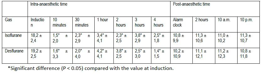
- Volatile anaesthesia: the team of Shepard et al. also showed a significant drop in tear production during gas anaesthesia. Even if TFS values rise above 10 mm/min on awakening, tear production remains reduced for at least 22 hours post-anaesthesia.
Finally, in a review published in 2022, Raušer et al. conclude that "higher doses of alpha2 adrenergic receptor agonists and combinations of anaesthetics, including inhaled anaesthetics, still significantly reduce tear production".
Protecting the cornea during sedation and anaesthesia
When there is a drop in tear production, the cornea is particularly vulnerable to epithelial erosion and even ulcers. To prevent this, it is therefore necessary to protect the ocular surface with a lubricant. In its recommendations "Anaesthesia and Monitoring of Dogs and Cats", the American Animal Hospital Association (AAHA) recommends "application of a corneal lubricant to protect the eyes from corneal ulceration after induction and every 2 to 4 hours", as well as "reapplication of eye ointment every 2 to 4 hours during the recovery period until an adequate blink reflex is present."
Applying a solution of sodium carmellose, 1% sodium hyaluronate or 0.25% sodium hyaluronate to 25 non-brachycephaly dogs every hour during anaesthesia and every 4 hours thereafter, for 24 hours, prevented exposure keratopathy in 90% of the eyes. At the end of the study conducted by Di Palma et al, none of the 25 dogs developed corneal ulcers.
In conclusion, it is recommended that an eye protector be applied as soon as the sedation or anaesthetic protocol is administered. This protector should be applied regularly during the operation. Its use may be extended on awakening, particularly in animals at risk of dry eyes (brachycephalic animals, for example).
MP Labo news: a study published with Lacri+®
Ophthalmology is an important subject for MP Labo, and this month we invite you to (re)discover an open multicentre study designed to assess the benefits and tolerability of Lacri+® in the treatment of dry eye. This study is freely available in the Irish Veterinary Journal.
Protocol
- 19 dogs with 1 or more signs of dry eye (keratitis, conjunctivitis, chemosis, ocular discharge) and a Schirmer's test between 5 and 10 mm/min.
- 2 drops of Lacri+® in each eye, twice a day for 30 days
- Veterinary checks at D0, D15 and D30:
Global ocular score (GOS): each sign (conjunctivitis, ocular discharge, ocular irritation
frequency of palpebral blinking, and corneal opacification/pigmentation - corneal vessels) is assessed from 0 (no signs) to 3 (severe signs). The GOS is the sum of the individual scores
Tears: quantitative monitoring (Schirmer STT 1 test) and qualitative monitoring (tear film break-up time)
Results
- Significant reduction in GOS from 2 weeks (p < 0.0001): - 31.1% after 2 weeks of use, - 54.5% after 4 weeks
- Schirmer's test values: significant increase from 2 weeks (p < 0.0001). 60% of dogs had a Schirmer's test greater than 10 mm/min at 4 weeks.
- No change in tear film break-up time
- Owner satisfaction: 83.3% of owners think the bottle is "easy" to "very easy" to use.
Lacri+® helps to increase the secretion of the tear film and improve the comfort and quality of life of dogs with dry eyes.